Skip to main content
Scanning Electron Microscopy (SEM)
Sector: Extractive Industries
Electron Microscopy is an extremely versatile tool which allows the study of both morphology and material composition from virtually all areas of science and technology.
The benefits of SEM over conventional microscopy include very high resolution and greater depth of field at magnifications from x200 to x1,000,000.
Typical Applications

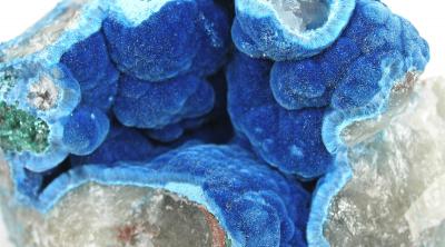
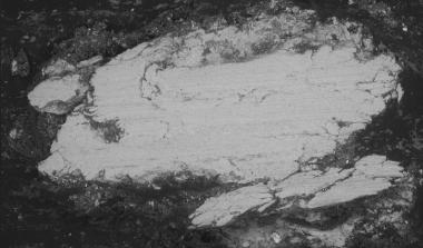
Mineralogy, grain size, mineral relationships, porosity.
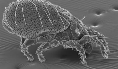
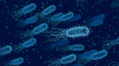
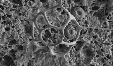
Plants, fungi, bacteria, insects.
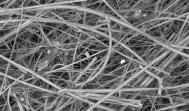
Glass, asbestos, natural, man-made.
The James Hutton Institute SEM lab is accredited for the analysis of metal chips and debris.
Instrumentation
- Carl Zeiss SIGMA VP Field Emission Scanning Electron Microscope
- Bruker Quantax 400 Energy Dispersive Spectrometer equipped with two Xflash 6/30 Silicon Drift Detectors
- Quorum Technologies PP2000T cryo-SEM preparation system
- Carl Zeiss Gemini 300 VP Field Emission Scanning Electron Microscope
- Bruker Quantax 400 Xflash FlatQuad Energy Dispersive Spectrometer
- Leitz Polarizing microscope equipped with a Spot ‘Insight’ digital camera (Diagnostic Instruments, Inc.
Techniques
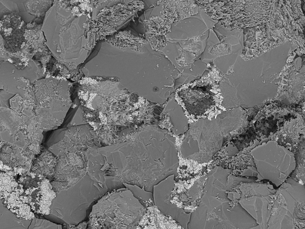
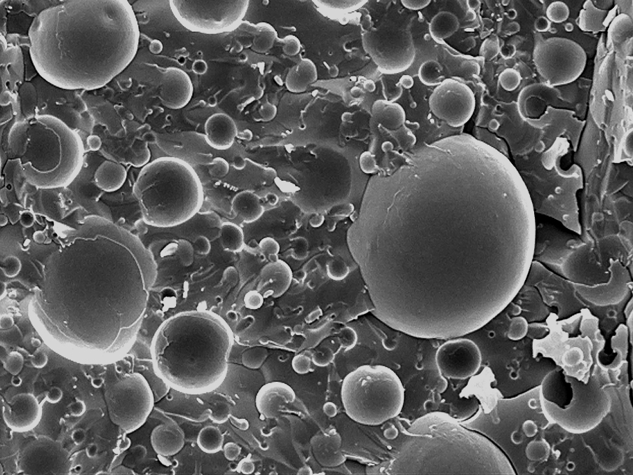
Secondary Electron Imaging
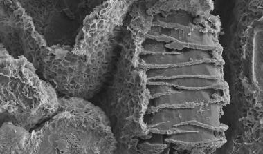
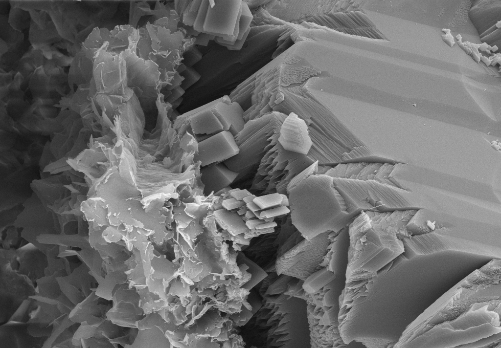 A beam of electrons is scanned across the surface of the specimen; images are built up from low energy secondary electrons which reflect the topography of the sample.
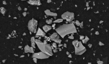
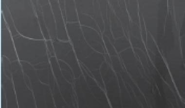
Backscattered Electron Imaging
 Backscattered electrons (BSE) provide an extremely useful signal for imaging in scanning electron microscopy as they respond to composition (atomic number or compositional contrast) and to local specimen surface inclination (topographic or shape contrast). BSE images obtained from flat polished surfaces reveal compositional changes due to variations in the average atomic number across the specimen.
X-ray Microanalysis
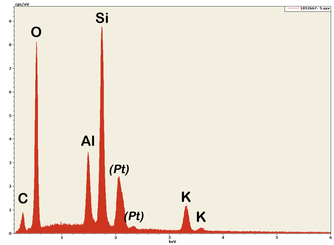 The electron beam is finely focused onto the specimen resulting in characteristic X-rays being produced from a microvolume (approximately 1µm3) of the sample. These X-rays are detected by an Energy Dispersive Spectrometer (EDS) and the results displayed as a spectrum. Each element has its own ‘fingerprint’ of peaks which allows both a qualitative and quantitative determination of the elements present in the selected region of the sample. EDS is an essential tool in geological applications combining elemental composition and morphology to identify minerals. It also has uses with biological specimens for example, localising elements such as calcium, potassium or phosphorus.
X-ray Mapping
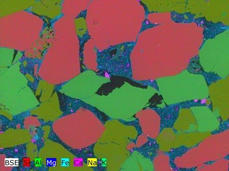 Digital element distribution maps can be collected simultaneously with electron image acquisition thus giving a visual representation of the chemical distribution in the sample. X-ray mapping is performed using HyperMap, a method whereby a complete spectrum is stored for each pixel of the acquired image. The collected database permits further processing of the data at any time during or after the measurement.
Cryogenic SEM
 Wet or liquid samples are cryogenically frozen to -170oC. An adapted SEM stage maintains the sample under cryogenic conditions during the analysis. Frozen samples can thus be analysed as solids using Secondary Electron Imaging, Backscattered Electron Imaging or X-ray Microanalysis.
Accreditation
The James Hutton Institute analytical laboratories operate to the standards required by UKAS accreditation and many of our routine techniques are accredited. A full accreditation schedule can be found at www.UKAS.com
Our ability to perform a huge variety of techniques means that in general a one-off analysis may not be accredited but our total commitment to high standards ensures it will be carried out to the exacting specifications that accreditation requires.
Contact us for further information.

Watch our free analytical webinar covering problem samples & scientific solutions / exploring investigative techniques for the energy sector
Case Studies
Other Services